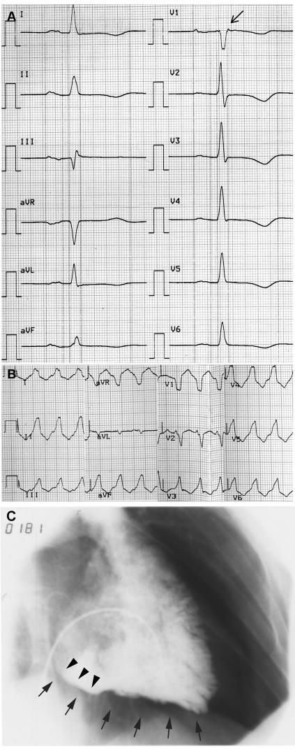
Figure 2. .
Clinical features of the mutation carrier. A, Twelve-lead resting ECG (50 mm/s). Note the T-wave inversions in the leads V1–V6 and the epsilon wave (arrow) in V1. B, ECG (25 mm/s) of a ventricular tachycardia with left bundle branch block morphology. C, Image of an angiogram showing an enlarged right ventricle in 30° right-anterior oblique, with evidence of diastolic bulging (arrowheads) at the right ventricle inflow tract and diffuse inferior hypokinesia (arrows).

