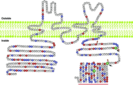
Figure 5. .
The predicted protein structure of TRIC-a, the longest isoform encoded by TRIC. Topology of the tricellulin was predicted by TMpred software. Of the 37 residues, 15 (40.5%) in the first extracellular loop are glycine and tyrosine. Red and blue amino acids indicate negatively and positively charged residues, respectively. The N- and C-termini consist of 31.6% and 41.4% charged residues, respectively. The position of an arginine codon mutated in family PKDF340 (p.R500X) is shown as a green R. Orange daggers indicate boundaries of the six coding exons of TRIC. Arrows indicate the locations of translation frameshifts due to splice-acceptor mutation (IVS3-1G→A) and splice-donor mutations (IVS4+2T→C and IVS4+2delTGAG), which occur at codons for residues C395 and K445, respectively. The red bar indicates the occludin-ELL domain.

