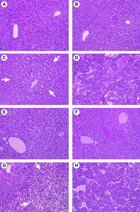
Figure 1.
Histology of the liver from DEN-injected and ASV mice. Histological findings in liver sections stained with hematoxylin and phloxin: A, normal liver in a 3-month-old noninjected male; B, liver in a 3-month-old DEN-injected male with no evidence of either architectural or cytological modifications; C, liver in a 6-month-old DEN-injected male showing a basophilic foci of altered hepatocytes (arrows); D, HCC in a 12-month-old DEN-injected male; E, normal liver in a 3-month-old control female; F, liver in a 2-month-old ASV male with hepatocellular atypias and mitosis; G, liver in a 3-month-old ASV male with a clear foci of altered hepatocytes (arrows) and atypias around the foci; H, HCC in a 6-month-old ASV male. Original magnifications, ×200.

