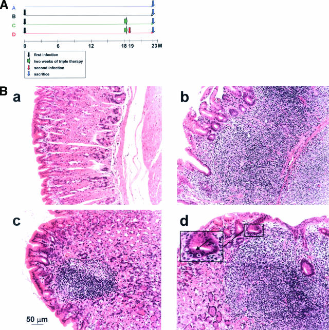
Figure 1.
Effect of Helicobacter eradication therapy and re-infection on MALT lymphoma progression. Groups of 8 to 12 mice were either infected for 23 months with Helicobacter felis (group B); infected for 18 months and then treated with triple antibiotic therapy (group C); or infected for 18 months, treated, and then re-infected with the same strain (group D). An additional group was left untreated (group A). At 23 months after initial infection, all mice were sacrificed, their stomachs were removed, and the tissue was processed for histological analysis. A: Schematic showing the timeline of treatments of all four groups. B: Representative H&E-stained sections of a control animal (a) and animals of groups B, C and D (b, c, and d, respectively). Animals of groups B and D are characterized by massive lymphocytic infiltration and the destruction of the glandular architecture of the gastric body mucosa. The arrow in the inset in d points to a typical lympho-epithelial lesion, a gastric gland that has been invaded by multiple lymphocytes. Gastric sections of animals in group C closely resemble control sections; however, roughly one-half of all successfully treated animals in this group retain small mucosal lymphoid aggregates (c), which are not accompanied by destructive lympho-epithelial lesions. Two of eight animals in group C were resistant to eradication therapy; gastric sections from these mice resemble those of groups B and D (not shown).

