Figure 5.
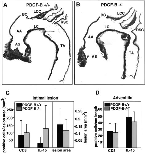
Detection of CD3- and IL-15-positive cells in longitudinal sections of ApoE−/−/PDGF-B chimeras. A, B: Representative aortic longitudinal sections stained with H&E from 45-week-old ApoE−/−/PDGF-B+/+ (A) and ApoE−/−/PDGF-B−/− (B) chimeras are shown. The location of the different vessels is shown: AS, aortic sinus; AA, ascending aorta; BC, brachiocephalic; LC, lesser curvature; LCC, left common carotid artery; RSC, right subclavian artery; TA, thoracic aorta. The brachiocephalic artery was removed where it branches off of the aorta and was embedded separately for analysis. C: Lesion T cells and IL-15-positive cells were evaluated by staining longitudinal sections of the thoracic aorta of 45-week-old chimeras (PDGF-B+/+, n = 8; PDGF-B−/−, n = 7) with anti-CD3 and anti-IL-15 antibodies, respectively. Lesion area is also shown for the two groups, and CD3- and IL-15-positive cells in intima were normalized to lesion area (mm2). D: CD3- and IL-15-positive cells in the adventitia were also counted and normalized to vessel length.

