
Fig. 7.
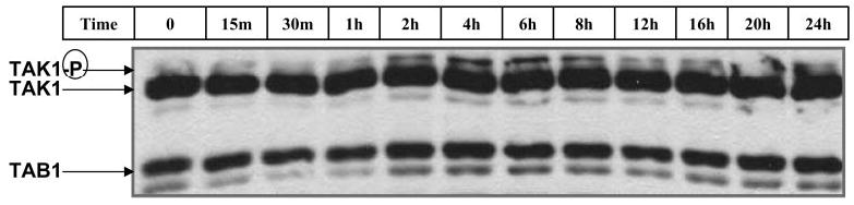
Activin phosphorylated TAK1 within 2 h and maintained TAK1 activation for 24 h. LβT2 cells were plated at 1 million cells per well in 6-well plates. Cells were pretreated with follistatin-288 (250 ng/ml; 16 h) and washed with culture media. Cells were then treated with activin (100 ng/ml) for 0, 15, or 30 min or 1, 2, 4, 6, 8, 12, 16, 20, or 24 h. Phosphorylation of endogenous TAK1 was detected by Western blot analysis as described in Materials and Methods.