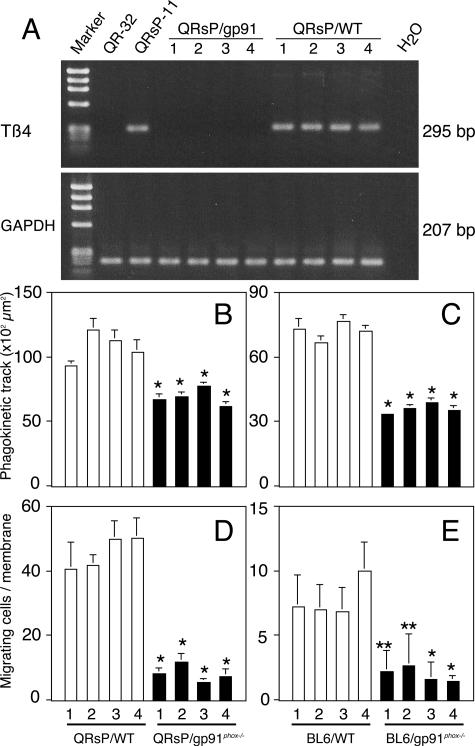
Figure 5.
Thymosin β4 gene expression and corresponding cell motility/invasion were augmented in tumor cells grown in WT mice but not those grown in gp91phox−/− mice. Tumor cells were established from QR-32 cells co-implanted with gelatin sponge in gp91phox−/− mice (QRsP/gp91phox−/−) and those in WT mice (QRsP/WT). B16BL6 cells were implanted into footpad in gp91phox−/− mice and in WT mice and established cell lines BL6/gp91phox−/− and BL6/WT, respectively. RT-PCR analysis for thymosin β4 and GAPDH (A), phagokinetic track patterns (B and C) and invasion through transwell (D and E) are shown. Each bar represents the mean ± SD of at least two independent experiments.*P < 0.001 and **P < 0.05, compared with tumors in WT with lowest motility or invasion.

