Figure 3.
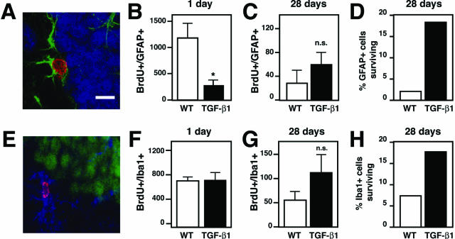
TGF-β1 mice have decreased astrogenesis, normal microgliogenesis, and prolonged glial survival. Coronal brain sections of TGF-β1 transgenic and nontransgenic littermate controls (n = 5 mice per analysis time point) injected at 8 weeks of age with BrdU to label dividing cells and sacrificed 1 or 28 days later for the presence of BrdU-positive astrocytes and BrdU-positive microglia. GFAP was used to label astrocytes, and Iba1 to label microglia. Bars are mean ± SEM. A: Confocal image of brain section containing a BrdU+/GFAP+ cell. BrdU, red; GFAP, green; and NeuN, blue. B and C: Quantification of the number of BrdU-positive astrocytes (BrdU+/GFAP+) 1 and 28 days after BrdU in TGF-β1 and their wild-type littermates. D: Percentage of the BrdU+/GFAP+ cells that remain 28 days later. E: Confocal image of brain section containing a BrdU+/Iba1+ cell. Iba1, blue; NeuN, green; and BrdU, magenta. F and G: Quantification of the number of BrdU-positive microglia (BrdU+/Iba1+) 1 and 28 days after BrdU in TGF-β1 and their wild-type littermates. H: Percent surviving BrdU+/Iba1+ cells at 28 days. *P ≤ 0.05, Student’s t-test. n.s., not significant. Scale bar = 10 μm.

