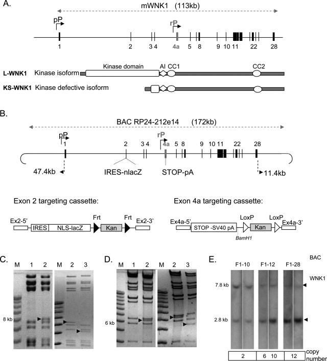
Figure 1.
Strategy for studying the production of WNK1 kinase domain-containing isoforms. A: Genomic structure of the mouse WNK1 gene. The genomic segment encompassing WNK1 is represented as a horizontal line, and exons are indicated by numbered vertical lines. The two alternative known promoters for mWNK1 are shown (bent arrows). The proximal promoters (pP) control transcription for the kinase domain-containing isoform (L-WNK1) and a renal promoter (rP) drives expression for the isoform lacking the kinase domain (KS-WNK1). AI, autoinhibitory domain; CC, coiled coil domain. B: Schematic diagram of the targeting cassettes used to generate the modified RP24-212e14 BAC and the resulting reporter BAC with the nlacZ gene inserted into exon 2 and a translation stop codon inserted into exon 4a, abolishing KS-WNK1 production. The size of the genomic sequences included in the BAC, on the 5′ and 3′ sides of WNK1, is indicated. The exon 2-targeting cassette was generated by inserting the nuclear lacZ gene, with the SV40 polyadenylation signal (nlacZ), downstream from an IRES sequence (internal ribosomal entry site). The kanamycin resistance gene (Kan) is flanked by two FRT sites (black arrows), which are recognized by the Flp recombinase. At each end of the cassette, sequences homologous to the 5′ (Ex2-5′) and 3′ (Ex2-3′) sequences of exon 2 facilitate recombination with the BAC. The exon 4a-targeting cassette was generated by inserting a transcription stop signal and the SV40 polyadenylation signal (STOP-SV40polyA), the kan gene flanked by two loxP sites (white arrows) and sequences homologous to exon 4a at each end (Ex4-5′ and Ex4a-3′). C: Exon 2 modification in the BAC was confirmed by EcoRV digestion. The exon 2 band displayed an appropriate increase in size and a decrease in size consistent with the deletion of the kan gene after recombinase treatment. D: Exon 4a modification in the BAC was confirmed by BamHI digestion. The fingerprint displayed an appropriate 6-kb band, confirming stop codon insertion and a decrease in size after recombinase treatment consistent with kan removal. Diagnostic changes are indicated by arrows. Lane 1: Parental BAC; lane 2: recombinant BAC with kanamycin resistance; lane 3: recombinant BAC with kan removed by Flp or Cre induction. E: The BAC transgene copy number was quantified by Southern blotting of DNA from F1 mice digested with PstI and probed with an ex4a probe. The expected size of the endogenous WNK1 gene is 7.8 kb and that for the transgene is 2.8 kb.

