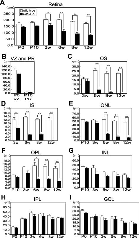
Figure 3.
Chronological changes of retinal degeneration as assessed by thickness of each layer at different ages in wild-type and Uchl3-deficient mice. A: Total retinal thickness is progressively decreased after 3w of age. B: Thickness of ventricular zone at P0 and photoreceptor layer at P10 shows no significant changes between both genotypes. C–F: Thickness of outer retinal layers in wild-type and Uchl3-deficient mice at different ages. The earliest change is revealed at 3w of age in inner segment of mutant retina (D). Thickness of outer segment (C), outer nuclear layer (E), and outer plexiform layer (F) in Uchl3-deficient mice is significantly decreased with age compared with that in the wild-type. G–I: Thickness of inner retinal layers in wild-type and Uchl3-deficient mice at different ages. Thickness of inner nuclear layer (G), inner plexiform layer (H), and ganglion cell layer (I) are unchanged between both genotypes. Each value represents the mean ± SE (*P < 0.05; **P < 0.01). In all panels, the white bars represent the thickness in wild-type mice and the black bars represent the thickness in Uchl3-deficient mice. VZ, ventricular zone; PR, photoreceptor; OS, outer segment; IS, inner segment; ONL, outer nuclear layer; OPL, outer plexiform layer; INL, inner nuclear layer; IPL, inner plexiform layer; GCL, ganglion cell layer.

