Figure 4.
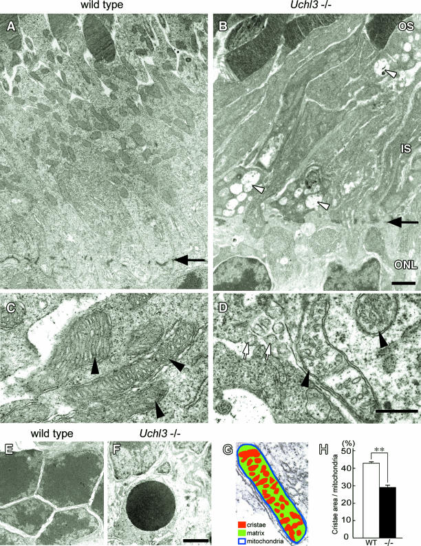
Ultrastructure of the outer retina in wild-type (A, C, and E) and Uchl3-deficient mice (B, D, and F) at 3w of age. A and B: Inner segment of mutant retina is shrunken associated with vacuolar changes (arrowheads in B). Arrows in A and B indicate outer limiting membrane. C and D: Subsets of mitochondria at the inner segment in Uchl3-deficient mice are swollen with decreased cristae (arrowheads in D) compared with that of wild-type (arrowheads in C). Groups of small round-to-oval shaped structures are occasionally seen in degenerated inner segment (white arrows in D). E and F: Outer nuclear layer of wild-type (E) and Uchl3-deficient (F) mice. Chromatin condensation of photoreceptor cells is observed in mutant mice (F). G and H: Morphometric analysis of mitochondria was performed with the percentage of cristae area (G; red) against mitochondrial area (n = 50 for each genotype). Cristae area in the inner segment is significantly decreased in mutant retina (H, −/−, black bar) compared with that in wild-type (H, WT, white bar). Each value represents the mean ± SE (**P < 0.01). OS, outer segment; IS, inner segment; ONL, outer nuclear layer. Scale bars = 1 μm (A and B), 500 nm (C and D), and 1 μm (E and F).

