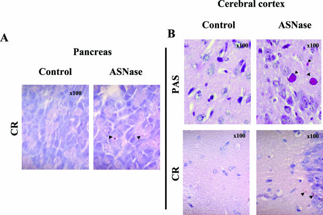
Figure 6.
Intracellular accumulation of proteins in pancreatic acinar cells (A) and frontal cortex white matter (B) from mice treated with ASNase (2 days with 10,000 IU/m2), as revealed by Congo Red and PAS staining under light microscopy. Arrowheads indicate β-amyloid (A, B) and glycoprotein (B) deposits. Original magnifications, ×100.

