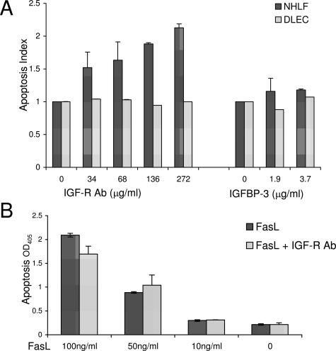
Figure 6.
Increased apoptosis of primary normal human lung fibroblasts treated with IGF-IR antibody. A: Normal human lung fibroblast or distal lung epithelial cells were serum-starved overnight and then incubated with indicated concentration of IGF-IR antibody or IGFBP-3 overnight. B: A549 cells were serum-starved and incubated with IGF-IR antibody (136 μg/ml) with or without the indicated concentration of Fas ligand. Apoptosis was measured by Cell Death ELISA-plus ELISA (Roche Applied Science) per the manufacturer’s directions. All experiments were done in triplicate and repeated at least twice. Apoptosis index is defined as the ratio of experimental condition OD405 nm:control (media alone) OD405 nm.

