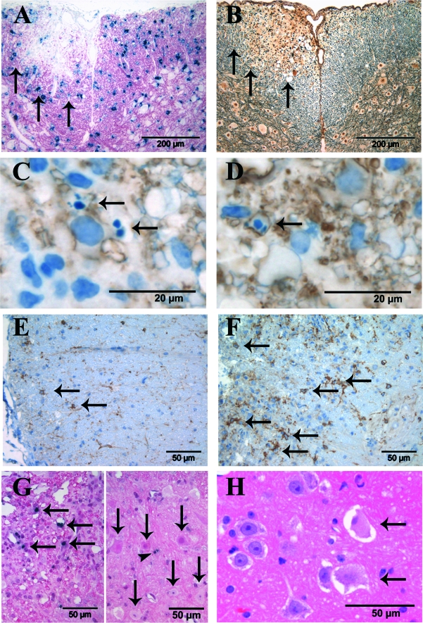
Figure 5-6916.
Morphological changes in EAE lesions. Immunohistological analysis of mice in the chronic disease phase (days 28 to 36 after immunization with MOG(35-55) peptide. A: Immunization with MOG(35-55) peptide leads to focal loss of oligodendrocytes and myelin [immunohistochemistry for PLP (red) combined with in situ hybridization for PLP mRNA (blue cytoplasmic signal)]. The arrows mark the border of the lesion. B: EAE lesions are characterized by a clear reduction in the number of axons (same lesion as in A, Bielschowsky’s silver impregnation). C: In EAE lesions cells with morphological characteristics of apoptosis such as condensed and/or fragmented nuclei are found (arrows). D: A proportion of apoptotic cells are oligodendrocytes as identified by immunohistochemistry with CNPase (brown). The apoptotic cell expresses CNPase on the cell surface (arrow). C and D: Immunohistochemistry with anti-CNPase, counterstained with hematoxylin. E and F: Immunohistochemistry for NG2 (brown) reveals a marked up-regulation of NG2-positive cells (arrows) in EAE lesions (F) compared to unaffected white matter (E). G: Within white matter lesions numerous cells with fragmented DNA are observed that stain positive in the TUNEL reaction (left, black arrows). The right panel depicts the adjacent gray matter. No TUNEL-positive neurons are seen in the gray matter (arrows), but one small nonneuronal cell is TUNEL-positive (arrowhead) [TUNEL staining (black) combined with nuclear fast red]. H: In the anterior horns of spinal cord gray matter swollen, weakly eosinophilic neurons were found that lack Nissl bodies (arrows).

