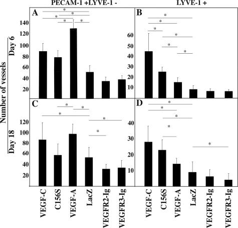
Figure 3.
VEGF-C enhances angiogenesis and lymphangiogenesis in the wound bed. A–D: Quantitative analysis of PECAM-1+ and LYVE-1− blood vessels and LYVE-1+ lymphatic vessels in the dorsal wound sections on days 6 and 18. Bars represent mean values ± 1 SD (n = 8). P values less than 0.05 are marked by an asterisk.

