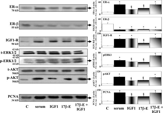
Figure 5.
Effect of serum, IGF-1, 17β-estradiol, and 17β-estradiol and IGF-1 on the protein expression (Western blot) of ER-α, ER-β, IGF-1R, ERK1/2, AKT, and PCNA in HuH-28 cell lines. HuH-28 cells deprived of serum for 48 hours were maintained under serum-deprived conditions for an additional 48 hours (controls, C) or exposed (48 hours) to serum, 17β-estradiol (17β-E, 10 nmol/L), IGF-1 (10 ng/ml, 1.3 nmol/L), or 17β-estradiol and IGF-1, which were added into the culture medium. For Western blot analysis, cells were solubilized in lysis buffer, and then the cell extract was resolved by 10% SDS-PAGE. The protein mass was determined by evaluating the intensity of the bands by scanning video densitometry and expressed (Prot. Expr.) as arbitrary densitometric units (A.U.) normalized to β-actin expression (ie, tested protein/β-actin × 100). *P < 0.01 versus controls (C); §P < 0.01 versus controls, P < 0.05 versus serum or 17β-estradiol and IGF-1. Ten independent experiments were performed for each protocol.

