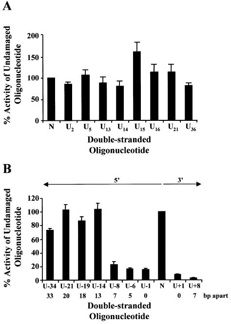
Figure 2.
Repair of uracil DNA damage in wild-type bacteria (BW35) Double-stranded oligonucleotides containing a single uracil (A), two closely opposed uracils (B) or no damage (represented as N on the graphs) were ligated into p3′luc and the ligation reaction co-transformed with either pPROLar.A122 or pACYC184 into wild-type bacteria. After a 4 h incubation, the luciferase activity was measured in a cell-free extract and the activity normalized to the number of kanamycin- or chloramphenicol-resistant colonies obtained after overnight growth on solid medium. These results were used to determine the percentage of activity measured for each sample compared with the undamaged control sequence. Triplicate transformations were performed for each ligation reaction and at least two ligations were examined. The average and the standard error are shown graphically. For substrates containing two uracils (B), the number of base pairs (bp) separating the two uracils, as well as the orientation (5′ or 3′) of the uracils with respect to each other, is indicated.

