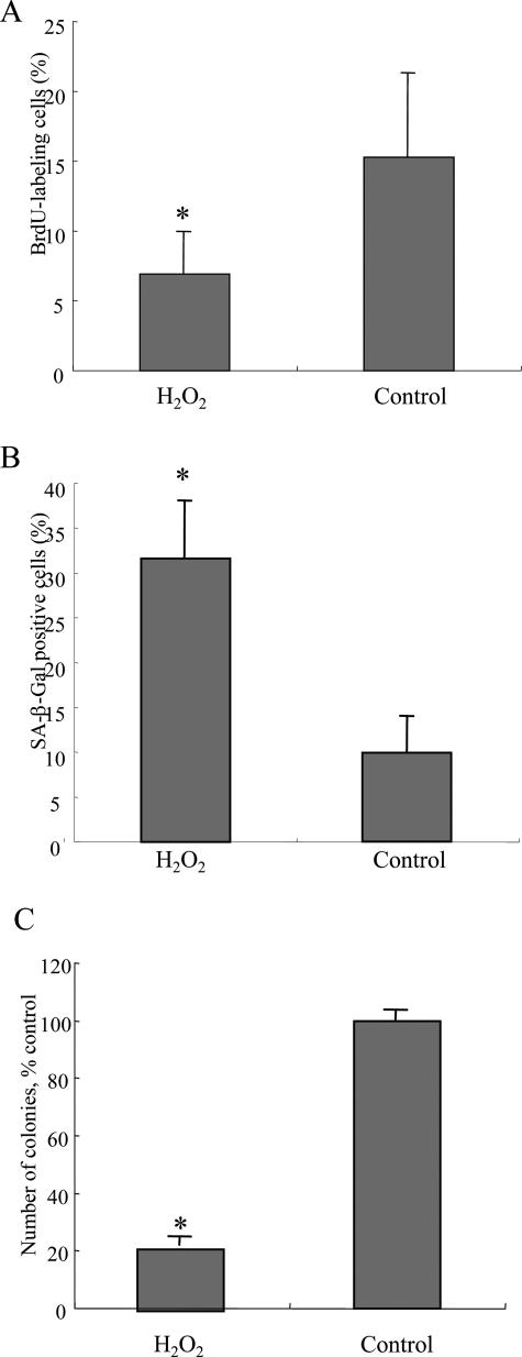
Figure 3.
Oxidative stress inhibits cell proliferation and induces cellular senescence. A: Cell proliferation was assessed by 5-bromo-2′-deoxyuridine (BrdU) assay on day 4 after H2O2 treatment. The labeling index of BrdU was counted for at least 1000 cells in each group. The labeling index of BrdU was significantly low in BECs treated with H2O2 (112.5 μmol/L, 2 hours), when compared with control BECs. Data are expressed as the mean ± SD. *P < 0.01 compared to control. B: Cellular senescence was assessed by senescence-associated β-galactosidase activity (SA-β-gal) on day 6 after H2O2 treatment. Percentage of cells positive for SA-β-Gal activity was significantly higher in BECs treated with H2O2 (112.5 μmol/L, 2 hours), when compared with the control BECs. Data are expressed as the mean ± SD. *P < 0.01 compared to the control. C: Cellular senescence was assessed by colony-forming assay on day 6 after H2O2 treatment. Number of colonies was significantly lower in BECs treated with H2O2 (112.5 μmol/L, 2 hours), when compared with the control BECs. Data are expressed as the mean ± SD. *P < 0.01 compared to the control.

