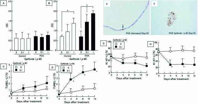
FIGURE 2.
Induction of apoptosis and inhibition of cell proliferative activity in cultured rat BECs by gefitinib. A and B: Apoptosis was determined using the ssDNA apoptosis enzyme-linked immunosorbent assay kit on day 1 (A) and day 3 (B) after gefitinib treatment (0 to 1 μmol/L). C and D: BEC apoptosis determined by the TUNEL method. The TUNEL-labeling index (LI) was determined in sections of collagen gel matrix of the three-dimensional cell culture as described in Materials and Methods (C, control rats; D, PCK rats). E and F: Immunostaining using anti-ssDNA antibody. Sections of collagen gel matrix of the three-dimensional cell culture were used. A few nuclei of biliary cysts were labeled with the anti-ssDNA antibody in PCK rats without gefitinib treatment (E, arrow), whereas many positive nuclear signals were observed with the disintegration of cyst morphology in the PCK rats treated with gefitinib on day 15 (day 8 after gefitinib treatment) (1 μmol/L) (F). G and H: Proliferative activity of BECs. The Ki-67-LI was determined in sections of collagen gel matrix of the three-dimensional cell culture as described in Materials and Methods (G, control rats; H, PCK rats). Data represent the mean ± SD in six sets (A and B) and three sets (C, D, G, and H). *P < 0.01. Original magnifications, ×400.

