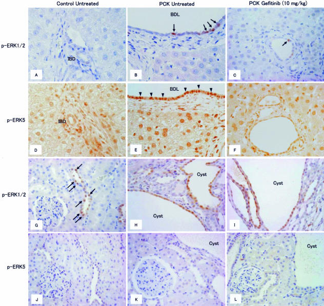
FIGURE 7.
Immunohistochemical analysis of the expression of phosphorylated (p-)ERK1/2 and p-ERK5 in the liver (A–F) and kidney (G–L). A few positive signals of p-ERK1/2 were observed in nuclei of biliary epithelium of PCK rats without treatment (B, arrows). Gefitinib (10 mg/kg) reduced the number of p-ERK1/2-positive nuclei of biliary epithelium of PCK rats (C, arrow). Increased expression of p-ERK5 was observed in biliary epithelium of PCK rats without treatment (E, arrowheads), and gefitinib (10 mg/kg) reduced the signal intensity of p-ERK5 (F). Positive nuclear signals for p-ERK1/2 were observed in the renal tubules of the control rats (G, arrows), and collecting tubule-derived cyst epithelium of PCK rats without treatment showed diffuse positive staining (H). I: Gefitinib (10 mg/kg) had no effect on the frequency and distribution of p-ERK1/2-positive cells in the PCK kidney. There were no p-ERK5-positive signals in the kidney (J–L). IBD, interlobular bile duct; BDL, bile duct lumen. Original magnifications, ×400.

