FIGURE 7.
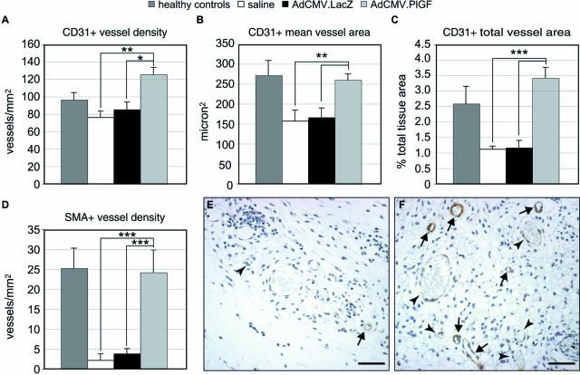
Analysis of granulation tissue vascularization. Central sections of wounds collected 7 days after wounding (6 days after treatment) were stained with an anti-PECAM/CD31 antibody or an anti-α-smooth muscle actin (SMA) antibody to detect endothelial cells and pericytes/smooth muscle cells, respectively. Vessel density (A, D), mean vessel area (B), and total area covered by vessels (C) were measured by computer-assisted morphometric analysis. In AdCMV.PlGF-treated diabetic specimens, total blood vessel density (A), mean vessel area (B), and the area covered by vessels (C) are significantly increased compared with both AdCMV.LacZ- and saline-treated diabetic wounds. In addition, the density of vessels surrounded by a continuous layer of SMA-positive cells is significantly increased in AdCMV.PlGF-treated wounds (D) (n = 6). Values are expressed as mean ± SEM. *P < 0.05; **P < 0.01; ***P < 0.005. The differences between AdCMV.PlGF-treated and healthy control wounds are significant only for the vessel density (P < 0.05). E and F: Representative fields of the granulation tissue of SMA-stained sections from AdCMV.LacZ-treated (E) and AdCMV.PlGF-treated (F) wounds, illustrating the increase in the density of “mature” vessels (arrows) in PlGF-transduced wounds. On the contrary, in AdCMV.LacZ-treated wounds the almost totality of vessels are not surrounded by a continuous layer of smooth muscle cells (arrowheads). Scale bars = 50 μm.

