FIGURE 7.
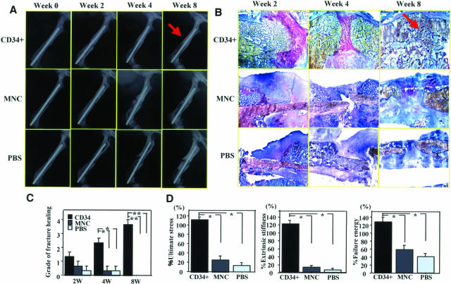
Radiographical, histological, and biomechanical evidence of fracture healing after CD34+ cell transplantation. A: Fracture healing was serially assessed by radiographs. By week 8, fracture was healed with bridging callus in all animals receiving CD34+ cells (arrow), but in no rats treated with MNCs or PBS alone. B: Serial assessment of fracture healing by histological examination with toluidine blue staining. Histological evaluation demonstrated the enhanced endochondral ossification consisting of numerous chondrocytes and newly formed trabecular bone at week 2, bridging callus formation at week 4, and complete union at week 8 (arrow) in animals receiving CD34+ cell transplantation. Although thick callus formation at week 2 was observed, the healing process had stopped by week 4, and the callus was finally absorbed at week 8 in animals receiving MNCs or PBS. C: The degree of fracture healing was assessed by Allen’s classification. The degree of fracture healing at weeks 4 and 8 was significantly higher after CD34+ cell transplantation compared with other treatments. *P < 0.05; **P < 0.01. D: Functional recovery after fracture is assessed by biomechanical three-point bending test at week 8. The percentage of all parameters (percent ultimate stress, percent extrinsic stiffness, percent failure energy) showing the ratio of each value in fractured side with contralateral side in animals receiving CD34+ cells was significantly superior to those in animals receiving MNCs or PBS. *P < 0.05.

