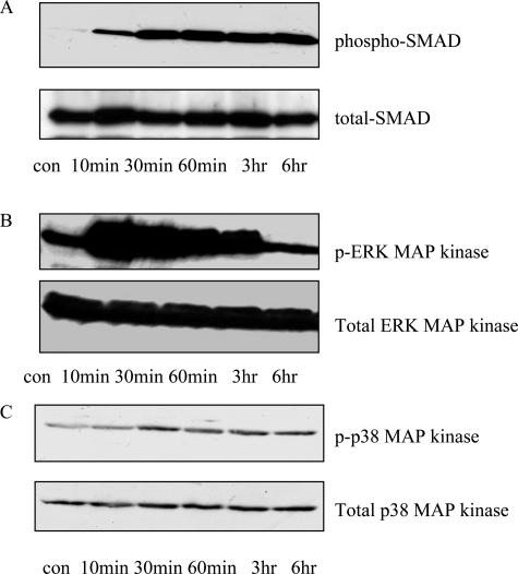
FIGURE 3.
TGF-β1 mediated activation of signaling pathways. Confluent monolayers of HK2 cells were stimulated by the addition of TGF-β1 (1 ng/ml) for up to 6 hours. At the time points indicated, total cell extracts were generated, and immunoblot analysis of lysate samples for phosphorylated-Smad/total Smad (A), phosphorylated ERK/total-ERK (B), and phosphorylated p38/total p38 (C) was performed.

