FIGURE 4.
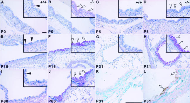
Goblet cell abnormal distribution in Hoxa5−/− lungs. Goblet cells were revealed by periodic acid/Schiff staining in postnatal lungs (A–J). At all ages tested, only a few positive cells were detected in wild-type samples (A, C, E, G, I; arrowheads), mostly scattered along the epithelium of the trachea (A, C) and the primary bronchi (E, I). No goblet cell was detected distally to the primary bronchi in controls (G). In Hoxa5−/− lungs, an increase in goblet cell number was systematically observed at every stage (B, D, F, H, J; open arrowheads), most obviously from P15 onwards (F, H, J). Furthermore, positive cells were detected in more distal structures such as the secondary bronchi (H) and bronchioles (not shown). Mucin MUC5AC, a marker of mucus hypersecretion, was detected by immunostaining (K, L). Although control samples remained negative (K), positive cells stained at their apical side were observed along the bronchi of Hoxa5−/− specimens from P15 onwards (shown for P31; L, open arrows). Scale bars = 50 μm.

