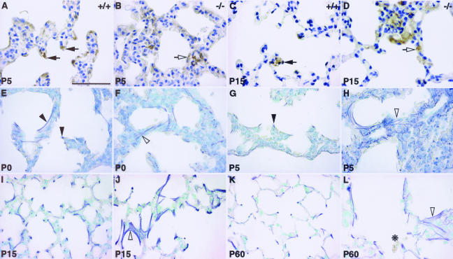
FIGURE 5.
Perturbed alveolar myofibroblasts localization (A–D) and elastin deposition (E–L) in Hoxa5−/− lungs. Alveolar myofibroblasts were located at the tip of the septa of control specimens at P5 and P15, as revealed by immunostaining with an α-SMA antibody (A, C; arrows). In Hoxa5−/− lungs, they were trapped within the parenchyma (B, D; open arrow). Weigert staining allowed visualization of elastic fibers (E–L). In P0 and P5 control specimens, elastic fibers were localized along the respiratory saccules and at the tip of the growing septa (E, G; arrowheads). In Hoxa5−/− lungs, elastin appeared tangled in the parenchyma (F, H; open arrowhead). At P15 onwards, Hoxa5−/− lungs displayed disorganized fibers (J, L; open arrowhead) compared with wild-type lungs (I, K). Moreover, inflammatory cells were detected in P60 mutant lungs (L, asterisk). Scale bar = 50 μm.

