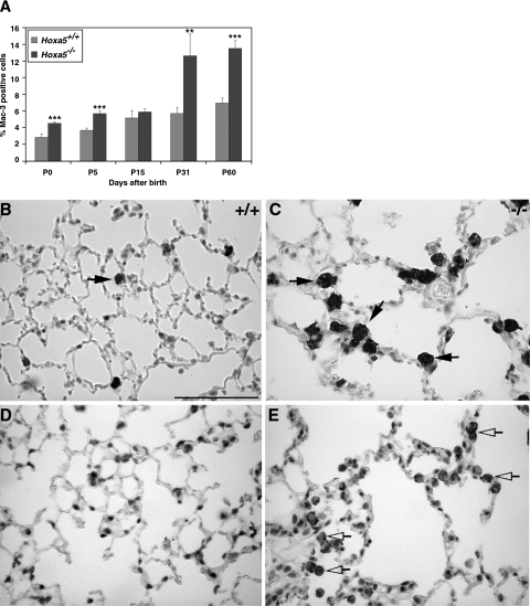
FIGURE 6.
Increased percentage of alveolar macrophages in Hoxa5−/− lungs. A Mac-3-specific antibody was used to detect macrophages in the lung (A–C, arrows) showing that the proportion of macrophages was statistically higher in Hoxa5−/− lungs than in controls. Asterisks denote statistical differences (**P < 0.01, ***P < 0.005). C: Moreover, at P31 (not shown) and P60, accumulation of large vacuolated macrophages was observed in Hoxa5−/− lungs. MMP-12 immunostaining revealed that the majority of macrophages in mutant samples produced this elastase (shown for P60; D and E, open arrows). Scale bar = 50 μm.

