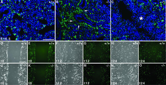
FIGURE 8.
Defective motility of alveolar myofibroblast progenitors. A–C: Visualization of the GFP-positive alveolar myofibroblasts progenitors in E16.5 wild-type lungs showed that progenitors were dispersed in the lung parenchyma (A). In contrast, most Pdgfαr/GFP-positive cells remained clustered around the epithelium in Hoxa5−/− lungs, indicating a block in distal spreading of the alveolar myofibroblast progenitors (B, arrows). Progenitors also failed to invade some lung regions (C, asterisk). Sections were counterstained with DAPI in blue. D–O: Wounding assays of primary mesenchymal cells isolated from lungs of E15.5 Hoxa5+/+;PdgfαrGFP/+ (D–I) and Hoxa5−/−;PdgfαrGFP/+ (J–O) embryos after 0 (t0; D, E, J, K), 12 (t12; F, G, L, M), and 24 hours (t24; H, I, N, O). Wild-type cells invaded the wounded area within 12 hours of the assay (F, G), filling the gap by t24 (H, I). In contrast Hoxa5−/−;PdgfαrGFP/+ cells showed reduced migration at both time points (L–O). Scale bars = 50 μm.

