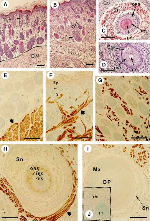
Fig. 2.
Histological views and localization of myosin in the skin and hair follicles detected by anti-skeletal muscle myosin antibody. Anti-myosin antibody clone A4 was used as the primary antibody in the immunohistochemical experiments. The results of hematoxylin and eosin staining are shown in A–D; A: dorsal skin, B: mystacial pad, C: a cross section of vibrissal follicle in the intermediate area and D: that in the upper (near-skin surface) area. The panels E–J represent the results of immunohistochemical staining; E: dorsal skin (the arrow denotes the dermal muscle arranged as a layer in the dermis). F, G: cheek skin (the arrow denotes the dermal muscle interwoven in the dermis of the mystacial vibrissal pad). H: a cross section of vibrissal follicle at the intermediate level. I: a cross section of vibrissal follicle at the level of the hair bulb. The arrow denotes the stained skeletal muscles surrounding the vibrissa follicle. J (inserted in I): a control sample of a section of vibrissal follicle processed without the primary antibody. In place of the antibody, the sample was treated with 1% bovine serum albumin. Sg, sebaceous gland; DM, dermal muscle; VF, vibrissal follicle; HS, hair shaft; IRS, inner root sheath; ORS, outer root sheath; Sn, sinus; Cs, capsule; Mx, hair matrix; DP, dermal papilla. Bars=100 µm.

