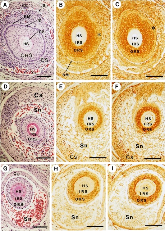
Fig. 5.
Co-localization of non-muscle myosin and actin in the outer root sheath. Bars=100 µm. Panels A, D and G show sections of vibrissal follicles stained with hematoxylin-eosin. Panels B, E, H and panels C, F, I represent immunostaining using anti-myosin antibody (BT561) and anti-actin antibody (C4) respectively. Each set of panels, (A, B, C), (D, E, F) and (G, H, I), consists of adjacent sections. HS, hair shaft; IRS, inner root sheath; ORS, outer root sheath; BM, basement membrane; DS, dermal sheath; Sn, sinus; Cs, capsule; *, the extension of the epidermal basal layer.

