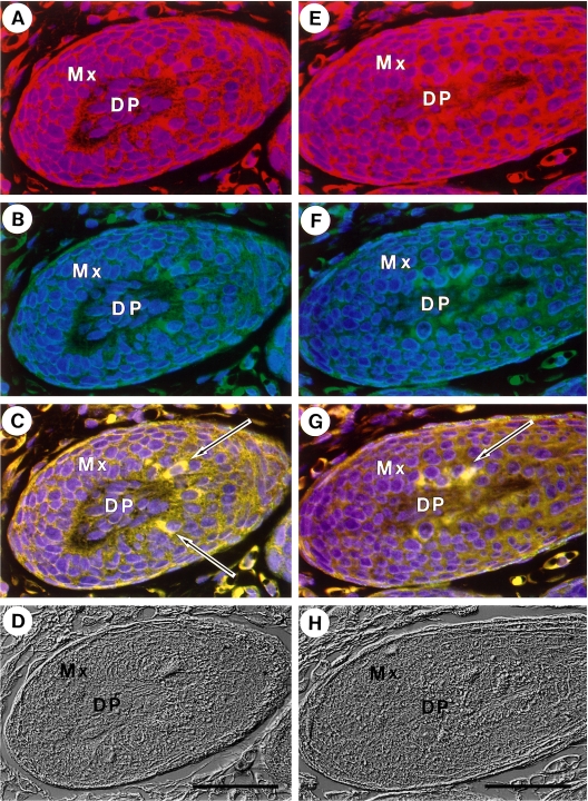
Fig. 6.
Co-localization of non-muscle myosin and actin in special hair matrix cells. Immunofluorescence staining. Panel A–D and panel E–H are the results obtained from a single section. Samples were stained for non-muscle myosin (red) (A, E) and actin (green) (B, F). Merged images are shown in C and G. Nuclei were stained with DAPI (blue). Corresponding Nomarski images are also shown (D, H). Bars=50 µm.

