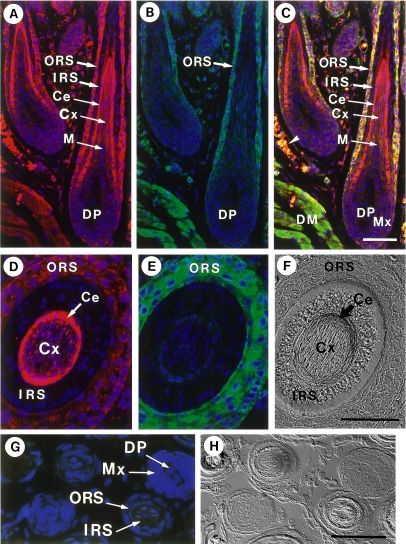
Fig. 7.
Immunofluorescence localization of non-muscle myosin in hair cuticle. Samples were stained for non-muscle myosin (red) (A, E) and actin (green) (B, F). Merged images are shown in C. Nuclei were stained with DAPI (blue). Corresponding Nomarski images are also shown (F). ORS, outer root sheath; IRS, inner root sheath; Ce, hair cuticle; Cx, hair cortex; M, medulla; Mx, hair matrix; DP, dermal papilla; DM, dermal muscle. A–C) A section of hair follicles prepared from the dorsal skin. An arrowhead in C designates autofluorescence of erythrocytes. D–F) A section of vibrissa follicle. G) Immunohistochemical control. A section was processed in the absence of primary antibodies. Merged images are shown in G). H) is a Nomarski image corresponding to G). Bars=50 µm.

