
Figure 7.
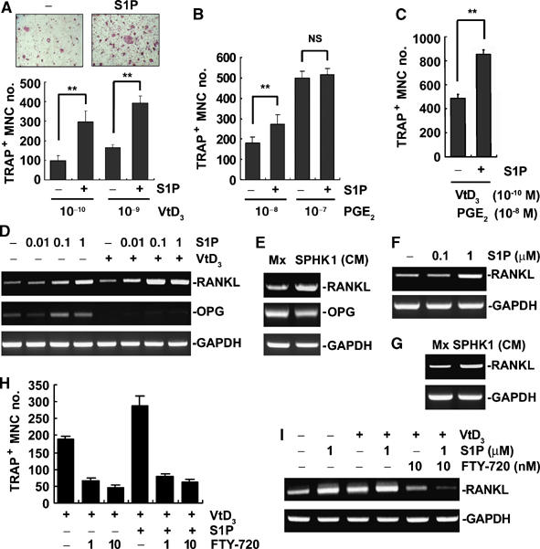
S1P potentiates osteoclast differentiation in coculture of BMMs and osteoblasts. (A) Coculture cells were treated with VtD3 and S1P (1 μM) for 6 days and TRAP+ MNCs generated were scored. **P<0.005 compared with S1P-untreated controls. (B) Cocultures were treated with PGE2 and S1P (1 μM) for 6 days. TRAP+ MNCs were counted. **P<0.005 versus S1P-untreated controls. NS, not significant. (C) Cocultures were carried out in the presence of PGE2 (10−8 M), VtD3 (10−10 M), and S1P (1 μM). TRAP+ MNC formation was assessed. **P<0.005 versus S1P-untreated controls. (D) Primary osteoblasts were treated with S1P and VtD3 (10−8 M) for 24 h. RANKL and OPG mRNA levels were analyzed by RT–PCR. (E) Osteoblasts were incubated for 24 h with indicated CM and RANKL mRNA levels were examined by RT–PCR. (F) T cells were activated with ionomycin and PMA and treated with S1P for 24 h. RANKL mRNA levels were determined by RT–PCR. (G) T cells were activated and treated with SPHK1-CM for 24 h. RT–PCR was performed to detect RANKL mRNA. (H) Coculture was carried out in the presence of FTY-720, VtD3 (10−9 M), and S1P (1 μM). TRAP+ MNCs were scored. (I) Osteoblasts were incubated with VtD3 (10−9 M), S1P, and FTY-720 for 24 h. RANKL mRNA was analyzed by RT–PCR.