
Figure 6.
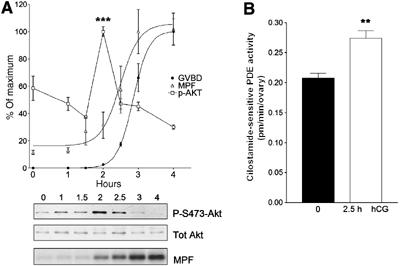
PKB/Akt and PDE3A activation precedes activation of the MPF complex. (A) Phosphorylation state of PKB/Akt at indicated hours after hCG injection was determined using Akt phosho-S473-specific antibodies (P-S473-Akt) and the amount of loaded protein was confirmed with Akt-specific antibodies (Tot Akt). Highly phosphorylated state of Akt is detected after 2 h from hCG injection. The value of p-Akt reports the ratio of phosphorylated Akt and total Akt (square). Activity of MPF complex (Cdc2/CyclinB) was measured with five randomly selected oocytes at each time point (triangle). The phosphorylation of histone H1, which is a substrate of MPF complex, started at 2 h after hCG injection and reached a maximum at 3 h (MPF). GVBD was counted by disappearance of the germinal vesicle or extrusion of the first polar body (filled circle). A representative of the three independent experiments performed is reported. *** represents P<0.005 compared to 0 h. (B) The mouse ovaries were removed and lysates immunoprecipitated with PDE3A antibody. The PDE3A activity was measured in the immune complex. After 2.5 h from hCG injection, 30% increase in PDE3A activity over basal was detected. The data are the mean±s.e.m. of three independent experiments. ** represents P<0.01 compared to 0 h.