
Figure 7.
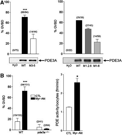
PDE3A is required for Akt-induced mouse oocyte maturation. (A) Oocytes from pde3−/− mice were injected with mRNA of WT PDE3A, M3–5, M1,2,6, or M1–6 mutant form. Vehicle (H2O) was injected as a control. Maturation of mouse oocytes was monitored by disappearance of the germinal vesicle or extrusion of the first polar body 19 h after mRNA injection. The injection of WT or M1,2,6 PDE3A mRNA induced meiotic resumption around 60–75% of oocytes, whereas about 30% of oocytes resumed meiosis when M3–5 or M1–6 mRNA was used. Numbers above the bars indicate the number of GVBD stage oocytes out of total injected oocytes. Protein expression of the injected mRNAs was compared with HA antibodies using 44 oocytes per each lane (left panel) and using 39 oocytes with PDE3A antibody (right panel). (B) Oocytes from WT and pde3a−/− mice were collected in M2 media, and WT oocytes maintained in meiotic arrest with 3.5 mM hypoxanthine. Left panel: Oocytes were injected with myr-Akt mRNA or H2O as a control. Injection of myr-Akt causes approximately 80% of meiotic resumption in WT oocytes but not in pde3−/− mouse oocytes. Right panel: Total PDE activity was measured with injected oocytes as described in Materials and methods. Approximately, two-fold increase in PDE activity was detected in myr-Akt-injected oocytes in three independent experiments. *** represents P<0.005 and * represents P<0.05 compared to control.