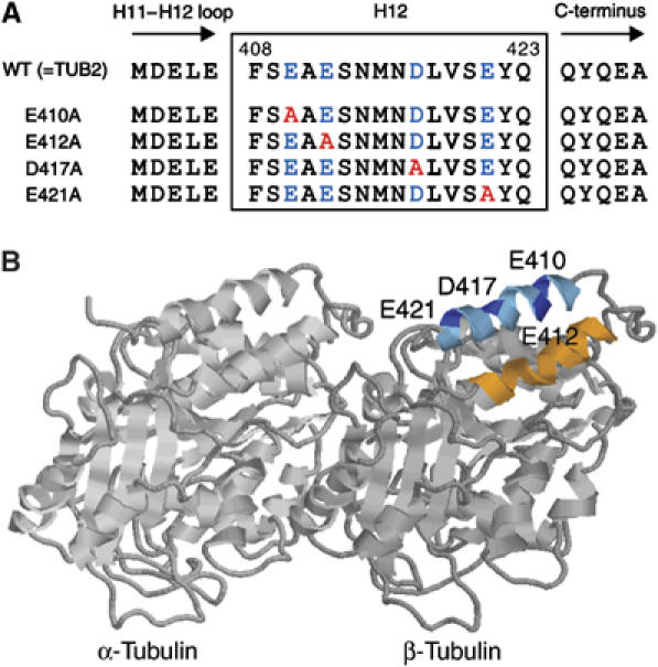
Figure 1.

Design of the H12 mutants of β-tubulin in Saccharomyces cerevisiae. (A) Sequences of the H12 region are shown with negatively charged residues indicated by blue and the residues substituted by alanines are indicated by red. (B) A ribbon diagram of the tubulin dimer viewed from the side of the microtubule with its minus end to the left (Nogales et al, 1998). Image analysis of the kinesin–microtubule complex revealed that in both nucleotide free and AMP-PNP state, kinesin motor domain is associated in close proximity to H11 (orange), H12 (cyan), and the COOH terminus (undefined in crystal structure) of β-tubulin (Kikkawa et al, 2000; Hoenger et al, 2000). The acidic residues in H12 mutagenized to alanine are indicated in blue.
