Figure 1.

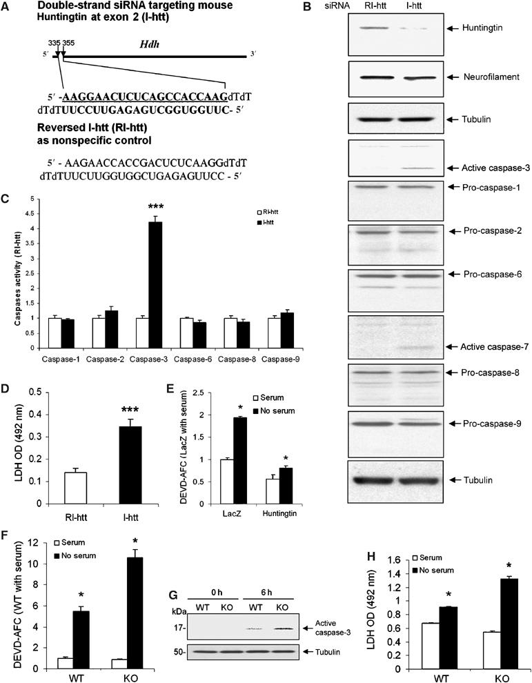
(A) Graphical representation of sites and sequences of double-strand siRNA targeting mouse huntingtin exon 2. I-htt represents the siRNA targeting huntingtin, and RI-htt represents reversed I-htt used as a control. (B) Endogenous huntingtin was depleted 72 h after transfection by I-htt in mouse neuroblastoma N2a cells. The blot was probed with an anti-huntingtin antibody (MAB2166). The same blot was stripped and then probed with antitubulin and antineurofilament H (200 kDa) antibodies to demonstrate equal loading. Densitometry of huntingtin signal was scanned and analyzed as ratio to tubulin. Huntingtin levels were 78.3±6.5% reduced by I-htt transfection when compared with N2a cells transfected with RI-htt. One hundred micrograms of same samples used for huntingtin blot were loaded to do immunoblots for caspase-1, -2, -3, -6, -7, -8, -9, and tubulin. Activation of caspase-3 and -7 was found after huntingtin reduction by siRNA. No activation of caspase-1, -2, -6, -8, and -9 was found. Immunoblots of tubulin demonstrated equal loading. The blots are representative of four independent experiments. (C) Caspase activation was evaluated using fluorogenic substrate assays. Caspase-3-like activity was increased 72 h after transfection by I-htt in N2a cells. Caspase-1, -2, -6, -8, and -9 like activities (YVAD-AFC, VDVAD-AFC, VEID-AFC, IETD-AFC and LEHD-AFC were used, in respectively, fluorogenic substrate assays) were not affected by huntingtin depletion. Data are expressed as the ratio of the I-htt group to the RI-htt group, n=3, ***P<0.001. One representative experiment out of five is shown. (D) Increased cell death was quantified by absorbancy values of LDH activity released into the medium 72 h after RI-htt and I-htt transfection. n=8, ***P<0.001. One representative experiment out of three is shown. (E) N2a cells cotransfected with CD19 and huntingtin or lacZ in a DNA ratio of 1:10. CD19+ cells were selected by magnetic beads coupled with anti-CD19. Overexpressing huntingtin inhibited caspase-3 activation in N2a cells after serum deprivation (n=3). (F) Following serum deprivation, Hdh−/− ES cells have increased caspase-3 activity (F, G) and cell death as detected by LDH release (6 h) (H) (n=6). All the above experiments were repeated at least three times, *P<0.05.
