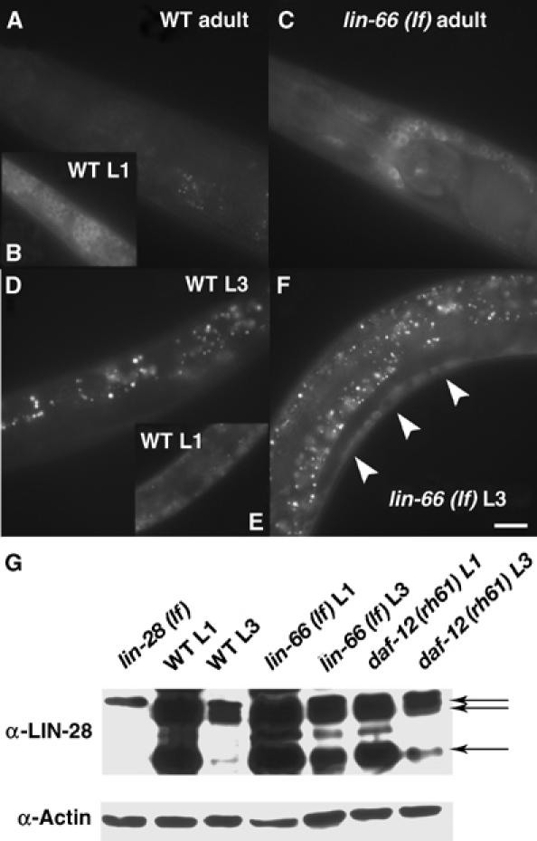
Figure 4.

lin-66 represses LIN-28 protein levels in late larval and adult stages. (A–F) Fluorescence images of animals of the genotype and stage as indicated. The fluorescence indicates the expression of an integrated lin-28∷GFP transgene (Moss et al, 1997). In L3 animals, strong expression is seen in Pn.p cell derivatives (cells above the white line) only in lin-66 (lf) mutants. In adults, the GFP expression is essentially undetectable in WT but still seen in many neurons in the mutant. Bar, ∼50 μm. (G) Western blot analysis of endogenous LIN-28 protein using an anti-LIN-28 antibody. Arrows indicate three LIN-28 protein bands determined in a previous study (Seggerson et al, 2002). A lighter exposure of the gel is shown in Supplementary Figure S3.
