
Figure 2.
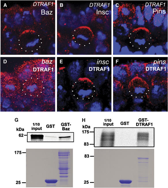
DTRAF1 interacts with Baz in vitro and its apical localization is dependent on Baz but not Pins and Insc. In DTRAF1 mutant embryos, Baz (red, A), Insc(red, B) and Pins (red, C) form crescents in the dividing NBs. The asymmetric localization of DTRAF1 (red) is dependent on Baz (D), but not Insc (E) and Pins (F). Cell body is outlined with dots. DNA is labeled in blue. Apical is up. GST-fusion protein pull-down assays suggest the possible physical interaction between Baz and DTRAF1 (G, H). The full-length GST-Baz fusion protein can specifically pull down the in vitro translated and 35S labeled full-length DTRAF1 (G). Similarly, the full-length GST-DTRAF1 specifically pulls down the in vitro translated and 35S labeled full-length Baz (H). The lower panels are the duplicated SDS gels stained with Coomassie brilliant blue, showing the molecular weights and quantities of recombinant GST, GST–Baz and GST-DTRAF1 applied on each lane. The diffused appearance of 35S labeled Baz is most likely due to the degradation of the in vitro translated Baz.