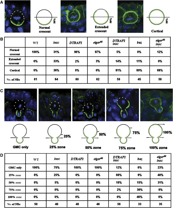
Figure 4.

Mira telophase rescue in various mutant backgrounds. The localization of Mira in metaphase NBs is subdivided into three classes: the normal crescents that occupy less than 50% of the NB cortex; the extended crescents that occupy more than 50% of the cortex and uniform cortical distribution, as indicated by the confocal images and the diagrams (A). Quantitation of Mira localization in metaphase NBs according to the standards set in panel A (B). Mira telophase rescue is quantitatively assayed by scoring of the ‘Mira tail' in four arbitrary zones (25, 50, 75 and 100%) according to its distance from the future GMCs as indicated by the confocal images and diagrams (C). The quantitation of Mira localization in telophase NBs following the criterion described in panel C is summarized in (D). NBs from stages 10/11 embryos are used. Apical is up. Mira is in green and DNA is in blue. White dots outline the cell body.
