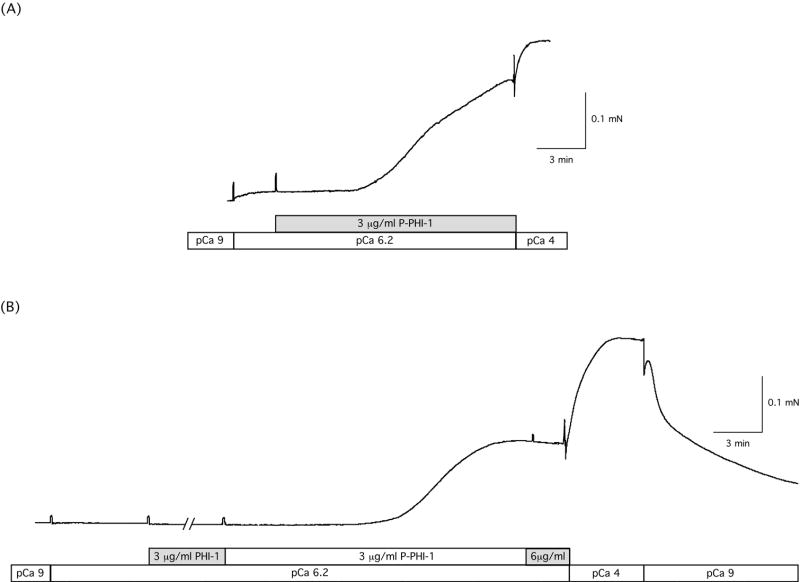
Figure 3. P-PHI-1 leads to Ca2+ sensitization.
(A) A skinned gizzard strip was placed in relaxing solution (pCa9) and then at the first transferred to pCa6.2. P-PHI-1 (3 μg/ml) was added to the pCa6.2 and the preparation was then transferred to pCa4 solution (without PHI-1). The pCa solutions changes and the addition of P-PHI-1 are indicated below the data trace. (B) A skinned gizzard strip was placed in relaxing solution (pCa9) and then transferred to pCa6.2. PHI-1 (3 μg/ml) and then P-PHI-1 (1st, 3 μg/ml and then 6 μg/ml) was added to the pCa6.2 solution. The fiber was then transferred to pCa4 solution (without PHI-1 or P-PHI-1) and then to pCa9 solution (without PHI-1 or P-PHI-1). The pCa solutions changes and the addition of PHI-1 and P-PHI-1 are indicated below the data trace. As is demonstrated, the addition of P-PHI-1, but not PHI-1, resulted in a significant increase in force.

