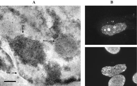
Figure 1.
Apoptin, expressed in human tumour cells, localises to heterochromatin and nucleoli. (A) Apoptin was detected in ultra thin cryosections of apoptin-expressing Saos-2 cells by immunogold staining. Indicated are: heterochromatin (HC), a nucleolus (NUC) and the nuclear membrane (NM); scale bar represents 200 nm. (B) Apoptin was detected in apoptin-expressing Saos-2 cells by indirect immunofluorescence microscopy. Top: Saos-2 cells, stained with anti-apoptin antibody 111.3. Bottom: nuclear morphology, as shown by DAPI staining. Similar results were published previously in Danen-van Oorschot et al. (3).

