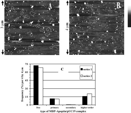
Figure 3.
Formation of higher-order MBP–apoptin–DNA complexes is highly cooperative. (A) MBP–apoptin combined with nicked pUC19 at a ratio of 11 kb per multimer, or four plasmid molecules per protein particle. In theory, one molecule can accommodate 25 multimers. Height is indicated in grey scale; bar is from 0.0 (black) to 1.5 nm (white). (B) MBP–apoptin combined with nicked pUC19 at a ratio of 3.8 kb per multimer. (C) Distribution of binding states of pUC19 molecules as observed in 18 separate SFM images, which were comparable with (B). Primary complexes correspond to DNA molecules with a single bound apoptin multimer; secondary complexes carry two multimers. Series 1 contains only higher-order complexes where DNA was clearly visible. Series 2 includes higher-order complexes of the expected size but without distinguishable DNA due to slightly different imaging parameters. There is no significant difference between both series.

