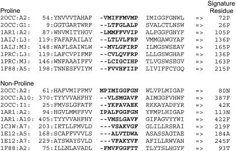
Figure 1.
Instances of proline- and non-proline-induced π-helical segments determined from the X-ray crystallographic structures of various proteins (PDB codes followed after the first colon by a letter designation for the protein chain and transmembrane helix numbering are indicated in the left most column). The residues N-terminal to the perturbing residue that form the π-helical segments are shown in bold, and the 10 residues on either side of the set of perturbing residues are shown with the numbering of the most N-terminal residue indicated on the left and that of the perturbing residue on the right.

