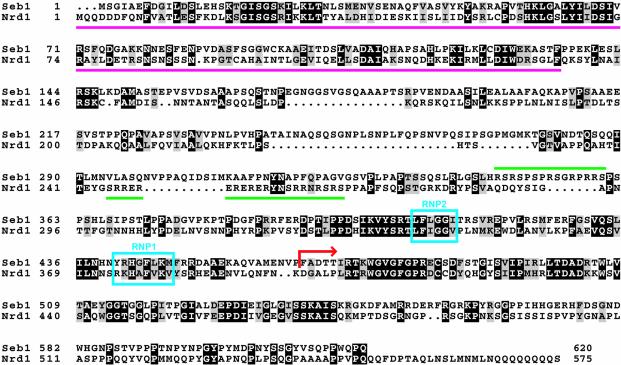
Figure 3.
Amino acid sequence alignment of S.pombe Seb1 and S.cerevisiae Nrd1. Identical residues are shaded in black and similar residues are shaded in gray. Magenta lines indicate the CTD-binding domain of Nrd1. Green lines indicate regions rich in arginine–serine or arginine–glutamate dipeptides. The RNP motifs are boxed in blue. A red arrow indicates the C-terminal region of Seb1 found to be fused to Gal4AD in pFb49.

