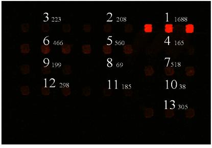
Figure 6.
Use of dendrislides to detect base mutations. Thirteen 20mer oligonucleotides were spotted in triplicate at 10 µM in Pi buffer 0.3 M, pH 9.0. These oligonucleotides corresponded to a part of the yeast HSP12 gene, and varied from each other by a single or two mutations proximal to the 5′- or 3′-end or in the middle of the sequence. The 20mer sequences from HSP12 were as follows: 1: NH2 5′-AATATGTTTCCGGTCGTGTC-3′; 2: NH2 5′-AATATGTTTCGGGTCGTGTC-3′; 3: NH2 5′-AATATGTT TCAGGTCGTGTC-3′; 4: NH2 5′-AATATGTTTCtGGTCGTGTC-3′; 5: NH2 5′-AATATGTTTCCGGTCGTGTA-3′; 6: NH2 5′-AATATGTTTCC GGTCGTGTG-3′; 7: NH2 5′-AATATGTTTCCGGTCGTGTT-3′; 8: NH2 5′-AATATGATTCCGGaCGTGTC-3′; 9: NH2 5′-AATATGGTTCCGG GCGTGTC-3′; 10: NH2 5′-AATATGCTTCCGGcCGTGTC-3′; 11: NH2 5′-AATAAGTTTCCGGTCGTGTC-3′; 12: NH2 5′-AATAGGTTTCCGGT CGTGTC-3′; 13: NH2 5′-AATACGTTTCCGGTCGTGTC-3′. Hybridisation was carried out with Cy5-labelled cDNA from yeast total RNA. At the top of the corresponding spots is given the mean value of fluorescent signal (intensity at 635 nm – background).

