Fig. 2.
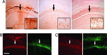
Neurotoxicity of serum containing anti-DNA, anti-DWEYS Ab. (A) (Left) Upon accessing the mouse brain after LPS treatment, we found that serum from an SLE patient with high-titered Abs to DNA and DWEYS bound CA1 neurons in the hippocampus, as revealed by anti-IgG staining. (Center and Right) Control serum lacking anti-NMDAR Ab (Center) and neurotoxic serum depleted of DWEYS reactivity (Right) were diffusely present but did not bind to CA1 neurons. (Scale bars: 800 μm; Inset, 100 μm.) (B) Neurons from mice given serum with anti-DWEYS reactivity and LPS treatment showed activated caspase-3 (red signal, Left) and fluorojade reactivity (green signal, Right). (Scale bar: 600 μm.) (C) Injection of purified IgG (2 mg in 100 μl of saline) from serum with high-titered anti-DNA anti-DWEYS Ab followed by LPS treatment reveals that the IgG bound hippocampal neurons and caused neuronal damage, as shown by activated caspase-3 (Left) and fluorojade (Right). Arrows identify the CA1 region of the hippocampus.

