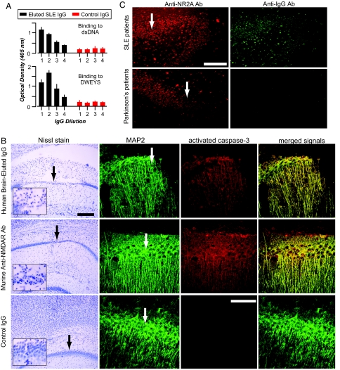
Fig. 4.
IgG with NR2A reactivity in postmortem brains of SLE patients. (A) The eluted IgG from lupus brain exhibits anti-DNA, anti-DWEYS reactivity. The graphs represent titrations of IgG binding by serial dilutions, starting at 5 μg/ml (dilution 1) and decreasing 2-fold each. (B) Eluted anti-DNA, anti-DWEYS Ab kills neurons. (Top and Middle) IgG (5 μg/2 μl) eluted from the brain of an SLE patient provokes widespread neuronal death when injected into the CA1 region of a mouse hippocampus (Top), similar to that of R4A, a murine monoclonal Ab that binds DNA and DWEYS (Middle). (Bottom) Injection of normal IgG causes no CA1 damage. (Scale bars: Top, 300 μm; Bottom, 150 μm.) MAP2 (green signal) labels neurons, and activated caspase-3 (red signal) identifies neurons destined to die. (C) Neurons in the hippocampus of SLE patients (n = 4) demonstrate colocalized immunoreactivity with Abs against the NR2A subunit (red signal) and human IgG (green signal). Hippocampal neurons from Parkinson's disease patients (n = 3) demonstrate immunoreactivity for the NR2A subunit but lack colocalizing endogenous Ab. Arrows identify the CA1 region of the hippocampus. (Scale bar, 300 μm.)

