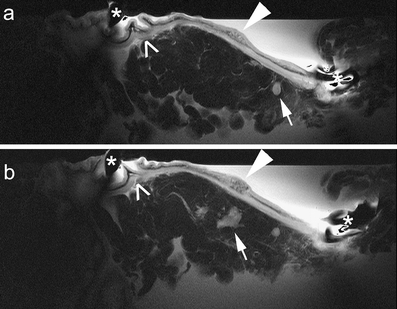
Fig. 2a, b.

Two consecutive T2-weighted images (a, b) of a T1 early gastric carcinoma (white arrowheads), well differentiated (intestinal type of Lauren classification), located at the subcardial region. Lymph nodes in adjacent fat tissue with a high signal intensity (white arrows) are visualized. Morphology of normal gastric wall is pointed out by open arrows. (*) marks the position of the receiver coil
