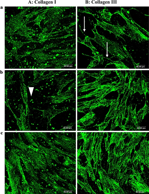
FIGURE 6.

Immunofluorescence staining of collagen in monolayer cultures of fibroblast on glass visualised by confocal microscopy. (A) Type I collagen is identified on the 3rd day of culture by use of anti collagen type I antibodies. Only spots of this type of collagen were secreted by the cells (arrowhead). (B) Type III collagen is identified by use of anti collagen type III antibodies. It appears like network in the cell (arrow). (objectif 40×/NA 0.80, Leica, Germany). Bar 40 μm. (a): Control cell (DMEM + 10%FBS); (b): Cell treated with 0.5 ng ml−1 TGF-β1; (c): Cell treated with 10 ng ml−1 TGF-β1.
