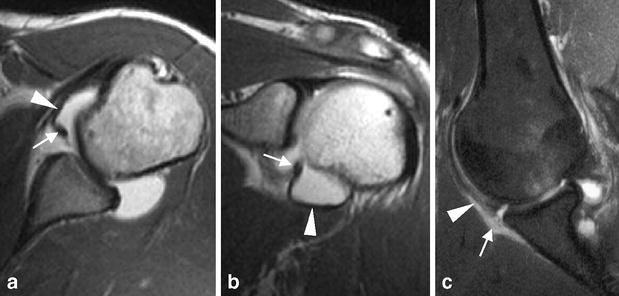
Fig. 3a–c.

Bankart lesion. a Transverse and b coronal oblique T1-weighted MR arthrograms show complete detachment of the anteroinferior labrum (arrows) which “floats” within the anterior capsular recess but remains attached to the IGHL (arrowheads). c Fat-suppressed T1-weighted MR arthrogram obtained in ABER position reveals separation of the anteroinferior labrum from the glenoid edge as well as disruption of the periosteum (arrow). Note taut IGHL adherent to the labrum (arrowhead)
